[gtranslate]

Benefits of IV Glutathione Therapy

16 Oct 2020

IV glutathione treatments, which help us have a glowing skin, help us get rid of toxins and heavy metals accumulated in our body, while also strengthening our immune system and increasing our resistance in the fight against diseases. While IV glutathione treatments applied at regular intervals regress the signs of aging on the skin, it also eliminates complaints such as chronic fatigue, widespread muscle pain and weakness, with its positive effects on the immune system and cell renewal, and helps us feel more vigorous and healthy.

Devamı

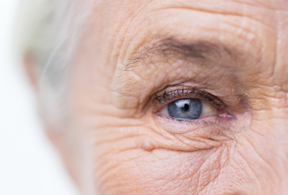
Aging Eye

17 Sep 2019

Today’s people are individuals in their 40s and 50s who are in an active working life, use computers extensively, and have to work at close range with their eyes for long hours.

Devamı

Beneficial Nutrients for Eye Health

15 Sep 2019

Let’s explore nutrient-rich food sources scientifically proven to be beneficial for our eye health, including vitamins A, E, and C, zinc, beta-carotene, lycopene, lutein, zeaxanthin, and other molecules with antioxidant properties.

Vitamin A and Its Significance for Vision Physiology

It is known that vitamin A plays a crucial role in visual physiology. To maintain good eye health, it’s essential to obtain an adequate amount of vitamin A through our diet. The rod cells in the retina are sensitive to vitamin A deficiency. Rod cells are responsible for our ability to see in dim light and for the rapid adjustment of our eyes to darkness. Studies have shown negative results in night vision threshold tests in individuals with low serum vitamin A levels. Prolonged vitamin A deficiency can lead to severe eye dryness, affecting vision in the cornea and conjunctiva. Animal-based sources rich in vitamin A include liver, fish oil, and egg yolk. Fatty fish such as salmon, tuna, and mackerel are especially rich in vitamin A and contain omega-3 fatty acids, which exhibit antioxidant properties.

Additionally, some of the carotenoids found in vegetables and fruits can be converted into vitamin A in the body. Analyzed carotenoids include alpha-carotene, beta-carotene, and gamma-carotene. Carotenoids are pigments responsible for the red, yellow, and orange colors in vegetables and fruits. Carrot, red pepper, mango, apricot, red grape, kiwi, and pumpkin are rich sources of beta-carotene. Some vegetables, although green in color due to the suppression of carotenoids by chlorophyll, are also rich in carotenoids. For example, broccoli and spinach, while green, are considered carotenoid-rich foods.

Lycopene has the highest degree of saturation with oxygen among carotenoids. Lycopene is a potent antioxidant, and foods such as tomatoes and watermelon are rich sources of lycopene.

Lutein and Zeaxanthin: Non-Vitamin A Carotenoids

Lutein and zeaxanthin are carotenoids that do not exhibit vitamin A activity. These carotenoids are predominantly found in plant-based sources. Vegetables like spinach, parsley, curly kale, broccoli, purslane, corn, and fruits like cantaloupe and oranges are abundant sources of lutein and zeaxanthin. These compounds act as potent antioxidants and are primarily found in the retina layer at the back of the eye. The retina layer experiences elevated oxidative stress due to light exposure, and lutein and zeaxanthin protect cells in the macula and retina from oxidative damage. Egg yolk is also a significant source of lutein and zeaxanthin from animal origin.

Vitamin E: Another Antioxidant

Vitamin E, like vitamins A and carotenoids, acts as an antioxidant. It exerts a protective effect at the cellular level by reducing oxidative damage caused by free radicals. Plant-based oils such as sunflower, corn, and soybean, as well as nuts like almonds, peanuts, and hazelnuts, are rich in vitamin E. Selenium, a mineral with antioxidant activity, works more effectively in the presence of vitamin E. Selenium is more prevalent in animal-based foods, including red and white meat, seafood, liver, and other organ meats.

Vitamin C: The Water-Soluble Antioxidant

Vitamin C is a water-soluble vitamin known for its antioxidant activity. Its advantage over other antioxidants is its ability to exhibit antioxidant effects in bodily fluids. Citrus fruits, green vegetables, rose hips, kiwi, tomatoes, and strawberries are rich sources of vitamin C.

Zinc: A Mineral for Eye Health

Zinc is a mineral found in abundance in muscles, liver, kidneys, and the retinal layer of the eye. It acts as an antioxidant by aiding the function of enzymes that reduce oxidative damage. Shellfish, especially oysters, are rich in zinc. Beef is also a source of zinc, while dairy products, though in lower amounts, contain this mineral. High consumption of fiber-rich foods can reduce serum zinc levels due to the binding of zinc by phytates in fibrous foods.

The Role of Antioxidants in Eye Health

Excess oxidative damage is known to accelerate the development of cataracts in adults. Consuming a diet rich in antioxidants is believed to slow down the formation of adult cataracts by protecting the lens from oxidative stress. Studies have also shown that the use of antioxidants can slow down the progression of senile macular degeneration (SMD), a retinal disease that typically occurs in old age.

Resveratrol: A Powerful Antioxidant

Resveratrol is a potent antioxidant that has gained attention in recent years. Red grape skins and seeds, as well as peanuts, are rich sources of resveratrol. It plays a crucial role in reducing oxidative damage and preventing premature cell aging.

Omega-3 Fatty Acids: Essential for Eye Health

Omega-3 fatty acids are molecules known for their antioxidant activity. They are essential, meaning they cannot be synthesized in the body and must be obtained through diet. DHA, an omega-3 fatty acid, is found in high amounts in the retina. It is believed to play a role in reducing oxidative stress in the retina. Additionally, recent studies have shown a connection between dry eye syndrome and omega-3 deficiency. Fatty, cold-water fish such as salmon, tuna, and mackerel are rich sources of omega-3. Among them, salmon stands out as an excellent source.

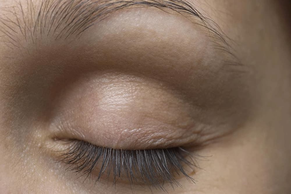
Eyelid Surgery

10 Sep 2019

Oculoplasty

Oculoplasty is a sub-branch of ophthalmology (the science of eye diseases). Oculoplasty deals with the diagnosis and treatment of bone and soft tissue diseases surrounding the eyelid, tear system and eye. The subject of oculoplastic surgery is the disorders related to the shape, function and appearance of the eyelids and their correction. Deformities in the eyelids may be congenital (such as congenital ptosis) or may develop in later ages due to factors such as familial inheritance and environmental factors. Aging alone causes changes in the shape of the eyelids and tissues around the eyes.

As a result of aging and the loss of collagen and elastic tissue in the skin combined with the effect of gravity, bagging and sagging of the eyelids, excess skin, drooping of the upper eyelid (ptosis) and drooping of the eyebrows are observed. In most patients, excess skin is accompanied by herniation of the fatty tissue around the eyes. Sagging of the lower eyelid may cause inward and outward turning of the lower eyelid edge. Excess skin and skin sagging on the lids can narrow the visual field and cause aesthetic concerns. In addition, wrinkles called ‘crow’s feet’ occur around the eyes due to the contraction effect of the muscle surrounding the eyelids.

What is blepharoplasty?

Excess skin and skin sagging that occurs on the eyelids with age is called ‘dermatochalasis’. It is usually more prominent in the upper eyelid and is often accompanied by fat herniation. In its treatment, eyelid surgery called ‘blepharoplasty’ is performed.

How to select the appropriate patient for blepharoplasty?

Before deciding on a surgical procedure, changes in the tissues around the eyes with age should be evaluated with a detailed physical examination. Is it the eyelid alone or the drooping eyebrow that causes the problem? Decision making is important in approaching treatment. In patients with prominent eyebrow ptosis, aesthetically pleasing results are obtained after eyebrow lifting, and blepharoplasty surgery can be postponed until later ages.

On the other hand, in patients where drooping eyebrows are the main problem, it should be taken into consideration that after upper eyelid surgery performed without considering eyebrow ptosis, the eyebrows will appear lower due to the decrease in the distance between the eyelashes and eyebrows and aesthetically pleasing results will not be obtained.

How should the patient who will undergo blepharoplasty be prepared for the operation?

Patients who will undergo blepharoplasty should be evaluated in terms of cornea, ocular surface diseases and dry eye during biomicroscopic examination. In addition, the patient should be questioned about allergies, the presence of systemic diseases that affect wound healing (such as diabetes), and the use of anticoagulant drugs that make bleeding control difficult.

What is xanthelasma?

Fat accumulation on the eyelid skin is called ‘xanthelasma’. Fat accumulation appears in the form of bright yellow plaques. These fatty plaques contain cholesterol. Its relationship with high blood cholesterol has not been fully elucidated, but it is more common in family members with familial high blood lipids. It should be considered that it may be associated with high serum cholesterol, especially when seen in young adults.

Xanthelasma is a common condition that usually affects both eyelids symmetrically. It is usually seen in middle-aged and older individuals. Its incidence is higher in women than in men. It is most commonly located on the part of the upper eyelids close to the root of the nose. Fatty plaques are formed by cholesterol-filled macrophage cells located in the upper layers of the dermis of the eyelid skin. It is not known exactly what causes this. However, in the presence of high serum cholesterol, relapses are observed more frequently after treatment.

Xanthelasma is not a malignant lesion. It does not cause clinical complaints; It does not affect visual function. Treatment consists of removing the lesions under local anesthesia for cosmetic purposes.

In the presence of large lesions affecting the entire lid, upper eyelid aesthetics (blepharoplasty) may be considered. Thus, better aesthetic results can be obtained by removing xanthelasma along with the excess skin tissue on the upper lid.

What is a mass on the eyelid?

The most common tumoral lesions in ophthalmology practice are eyelid masses. The majority of masses seen on the eyelid are benign tumors. The structure of the eyelids contains different tissues such as skin, mucosa, muscle tissue, sweat and sebaceous glands, and a wide variety of benign or malignant tumors can originate from these structures. Masses that appear on the eyelid can also be seen anywhere on the body covered with skin. However, the clinical appearance, histological features, and surgical principles applied in the treatment of these tumors are different from skin lesions seen in other parts of the body.

The most common benign valve tumor in adults is squamous cell papilloma. Irregular hyperkeratinized squamous epithelium

EYELID BROUGHTS, CAUSES AND SOLUTION SUGGESTIONS

29 Jul 2019

The skin on the eyelids is structurally very thin. The distribution of melanin pigments, which give color to the skin, determines the color of the skin on the eyelids.

Melanin is a natural pigment produced by special skin cells called melanocytes. Skin with a higher amount of melanin appears darker in color, while skin with lower melanin levels appears lighter. Additionally, the distribution of superficial blood vessels also affects the color of the skin, especially in thin-skinned areas like the eyelids.

When there is darkening of the skin in a specific area, it is referred to as hyperpigmentation. This occurs due to increased melanin production in that area. The causes of darkening around the eyelids, often associated with increased pigmentation, are typically related to various factors that occur in and around the eyes. It’s important to distinguish hyperpigmentation caused by increased pigmentation from discoloration that can result from artificial dyes or chemical burns. Hyperpigmentation-related color changes usually manifest as circular dark rings around the eyes.

Most often, darkening of the skin on the eyelids is due to inflammation in this area. Inflammation is a process initiated by the body to protect tissue from damage and is usually a response to infection or trauma. Inflammation is typically a short-term condition, but when it persists, it can stimulate melanocytes, leading to increased melanin production and skin darkening.

Allergies are an example of long-term and recurrent inflammation. People with allergic rhinitis or asthma often experience eye allergies as well. Prolonged allergic inflammation around the eyes can lead to darkening of the skin.

In cases of allergic conjunctivitis, another reason for skin darkening is trauma caused by repeated rubbing of itchy eyelids. Rubbing the eyelids can lead to skin darkening.

Furthermore, in allergic individuals, repeated applications of allergens such as cosmetics can lead to a skin reaction called allergic contact dermatitis. Contact dermatitis is an example of prolonged inflammation, and it can cause changes in skin color on the eyelids over time.

When exposed to various factors that cause damage, such as mechanical, chemical, or electromagnetic factors, permanent skin darkening can occur on the eyelids. Continuous rubbing of the eyelids can result in mechanical damage, while exposure to strong acids or similar substances causes chemical damage. Sunburn is an effect of electromagnetic damage, although skin darkening caused by sunburn on the eyelids is generally temporary.

Some substances, even if natural and harmless, like tears or sweat, can cause discomfort when exposed to the skin for extended periods. Such substances can lead to irritant contact dermatitis. Consequently, itching, redness, and a burning sensation can occur around the eyelids. Repeated rubbing of itchy eyelids due to itching can result in skin darkening.

A lack of sleep for extended periods can cause skin darkening around the eyelids. The exact cause of this phenomenon has not yet been fully explained. It is thought that inadequate sleep causes the blood vessels around the eyes to dilate, making the eyelids appear darker. Additionally, the dilation of blood vessels can lead to fluid leakage into the surrounding tissue, often resulting in both skin darkening and puffiness around the eyelids.

In cases of certain nutritional deficiencies, such as iron-deficiency anemia and B vitamin deficiencies, dark circles around the eyes can be observed.

Genetic factors, advancing age, and the use of cosmetic products are other factors that can contribute to skin darkening on the eyelids.

Here are some simple tips for reducing dark circles around the eyes:

  1. Cold Compress: Applying a cold compress to the eyelids is a known method to reduce inflammation. Placing an ice pack or even cucumber slices (chilled in the refrigerator) on the eyelids can provide relief due to their cooling effects.
  2. Get Adequate Sleep: Increasing the number of hours of sleep can help alleviate dark circles.
  3. Balanced Diet: Maintaining a well-balanced diet can have a positive impact on the overall health of your skin.
  4. Avoid Eye Makeup: In cases where there is swelling and itching along with eyelid darkening, it is advisable to avoid eye makeup.
  5. Use UV-Blocking Sunglasses: Wearing sunglasses with UV protection, especially on sunny days, can help prevent an increase in dark circles around the eyes.

When considering treatment for dark circles around the eyes, it’s essential to identify and address any underlying causes. Tailoring the treatment approach to address the root cause can enhance effectiveness and reduce the likelihood of recurrence. Since the skin on the eyelids is very thin, skin-lightening and exfoliating methods applied to other areas of the body may not be suitable for the eyelids. It is recommended to investigate various factors mentioned above, such as allergies or nutritional deficiencies, before starting any treatment, as this can contribute to post-treatment satisfaction.

HIGH CHOLESTEROLEMIA AND EYE HEALTH

29 Jul 2019

High blood cholesterol levels have observable signs in the eyes. High serum cholesterol levels can often lead to various cardiovascular problems, which are sometimes referred to as high cholesterol symptoms.

Devamı

EFFECTS OF THE SUN ON OUR EYE HEALTH

29 Jul 2019

Following a long and cold winter, we are finally embracing the joy of the much-awaited spring season and sunny days. During these days when we bask in the sun, I wanted to take a closer look at the effects of UV rays on our eye health.

Devamı

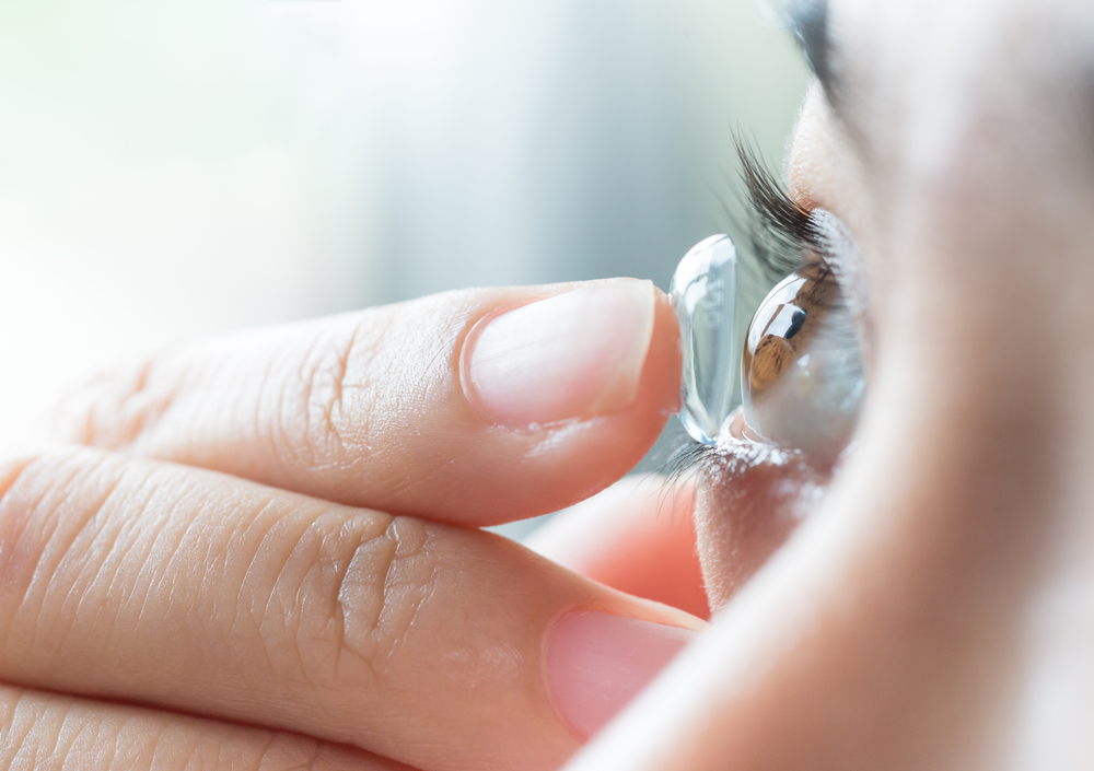
Basic Information About Contact Lenses

29 Jul 2019

“Contact lenses are medical products that are placed directly on the transparent layer of the eye called the cornea. They can be used for correcting refractive errors, preferably instead of glasses, or for cosmetic purposes such as changing eye color.

In addition to this, they are used as part of the treatment in some eye diseases such as keratoconus or in cases where the refractive errors between the two eyes are significantly different, especially in children.

Contact lenses can be classified based on the material they are made of:

  • Soft lenses are made from a type of plastic called hydrogel, which is a gel-like material that can hold water. They are soft and flexible, making them easy to use. They quickly adapt to the eye, so they do not cause discomfort. They are thin enough to allow oxygen to pass through to the cornea.
  • Silicone hydrogel lenses are an advanced type of soft contact lens. Compared to hydrogel lenses, they have a more porous structure and allow more oxygen to reach the cornea. They have been in use since 2002.
  • Classic hard lenses are made of a hard and transparent plastic material called PMMA. These hard lenses do not allow oxygen to pass through and are no longer in use.
  • Gas-permeable lenses can be considered as the new generation of hard lenses. These lenses are made by combining plastic with other materials such as silicone and fluoropolymers. They have a structure that allows oxygen to pass through. Since their introduction in 1978, they have replaced classic hard lenses. Due to their rigid structure, they provide clearer vision compared to soft lenses, but soft lenses are the most commonly preferred lenses today due to their ease of use and comfort.
  • Hybrid lenses are designed with the idea of combining the optical clarity provided by gas-permeable hard lenses with the comfort of soft lenses. They have a high gas-permeable hard section at the center of the lens, surrounded by lens sections made of hydrogel or silicone hydrogel material.

Contact lens designs are named according to their purpose in correcting vision problems:

  • Spherical contact lenses are typically round in design. They are used to correct myopia and hyperopia.
  • Toric contact lenses are used to correct astigmatism and can also address hyperopia and myopia associated with astigmatism.
  • Bifocal contact lenses contain separate optical zones for distance and near vision and are used to correct presbyopia.

Contact lenses have specific applications for cosmetic purposes. Colored lenses can be used to change eye color, and there are also colored prosthetic lenses used to cover aesthetic issues in traumatized eyes.

There are three fundamental parameters that determine the suitability of contact lenses for the eye:

  • The base curve, which determines the curvature of the lens’s back surface that sits on the cornea. It is usually expressed in millimeters (e.g., 8.4 mm, 8.5 mm).
  • The diameter of the lens, which represents the maximum length measured from one end of the contact lens to the other. Gas-permeable lenses typically have diameters in the range of 9.0 to 9.8 mm, while soft contact lenses have diameters ranging from 13.4 to 15.0 mm.
  • The power of the lens, which indicates the optical refractive power of the lens and is expressed in diopters (D). It represents the degree of refractive error in the eye.

When selecting the right contact lens for an individual, measurements of corneal diameter and refractive values of the eye are determined by an eye specialist. It should be noted that the lens prescription often differs from one’s glasses prescription.

WHEN USING CONTACT LENSES

Contact lenses are medical products that are placed directly on the cornea of the eye. The use of contact lenses became widespread with the introduction of soft contact lenses made of hydrogel, a gel-like material containing water, in the 1970s. Today, the most commonly used contact lenses are silicone hydrogel lenses, which are an improved form of soft lenses. These lenses allow more oxygen to pass through to the cornea compared to hydrogel lenses.

The cornea is the transparent front layer of the eye, resembling a bulging watch glass. It serves as a window for incoming light rays, effectively bending the light to focus it on the retina at the back of the eye. It is the most powerful refracting medium in the eye, and its transparency is maintained by being avascular. It consists of 78% water and the remaining part is collagen tissue. It maintains its transparency and shape through intraocular pressure. Oxygen and nutrients are supplied to it through tears and the atmosphere since it lacks blood vessels. To ensure that the corneal layer receives sufficient oxygen and nutrients, it is important to remove contact lenses when going to bed.

Extreme care should be taken when using contact lenses that make direct contact with the corneal layer. It should be remembered that incorrect usage can lead to permanent visual impairment due to damage to the corneal layer. When starting to use contact lenses and throughout their use, it is essential to be under the supervision of an eye specialist.

Contact lenses, when used carefully and correctly, are effective and safe in correcting refractive errors such as myopia, hyperopia, and astigmatism. They are preferred over glasses by young people and young adults because of their comfort and ease of use, making them suitable for active and fast-paced daily life. Additionally, contact lenses can be used for cosmetic purposes to change eye color in individuals without vision problems or as part of the treatment in certain corneal diseases like keratoconus.

There is no specific age to start using contact lenses. In medical cases, doctors can prescribe contact lenses even for infants and small children. When starting to use contact lenses, the fundamental principle is to have contact lenses prescribed by an eye specialist that match the individual’s corneal diameter and eyeglass prescription. To ensure the safe use of contact lenses for many years, it is important to follow the usage instructions recommended by the doctor and to remain under their care.

Contact lenses are safe medical products used by most people. However, the belief that everyone can use contact lenses is a misconception. In some cases, contact lens use may not be suitable for individuals. People who frequently experience eye infections, those with severe eye allergies, individuals with dry eyes, people who work in dusty and dirty environments, and those who do not have the ability to properly care for contact lenses are not suitable candidates for contact lens use.

The most feared complication in contact lens users is corneal infections that can lead to temporary or permanent vision loss. Risk factors that increase the chances of corneal infections are often related to usage errors. Daily disposable lenses should never be reused once they are removed from the eye. Monthly lenses should be removed every night, as sleeping with lenses increases the risk of eye infections. Monthly lenses should be replaced without delay, without extending their usage period. It should be remembered that prolonged use of lenses can increase the risk of eye allergies, eye infections, and reduce the comfort of lens wear due to the accumulation of proteins on the lens.

How should I care for my contact lenses?

After removing contact lenses from the eye, they should be thoroughly cleaned and disinfected with multi-purpose contact lens solutions.

The lenses placed in the solution after removal should be left in the solution for at least 5-6 hours and should not be taken out immediately and reinserted into the eye.

Contact lenses should be cleaned every day, and the lens

case should be replaced every three months.

After inserting the lenses into the eye, the solution in the lens case should be emptied, and the lens case should be left open to air dry.

Contact lenses should never be cleaned with any cleaning solution other than contact lens solution.

Except for the eye drops recommended by your doctor, no other eye drops should be used while wearing contact lenses.

What should I be cautious about when using contact lenses?

Hands should be washed thoroughly before touching contact lenses, and hands should be free of creams, lotions, or cosmetic residues.

Make sure that the lens is undamaged and well-cleaned before inserting it into the eye.

Lenses are personal to the individual and should never be shared.

Even for cosmetic purposes, lenses should be prescribed by an eye specialist after measuring the corneal diameter.

Contact lenses should be used in accordance with the schedule recommended by your doctor, and wearing them for extended hours should be avoided, as well as sleeping with lenses, as it increases the risk of eye infections.

Contact lenses should be replaced with new ones at the time recommended by your doctor. The duration of lens use may vary from person to person.

When the following conditions occur while using contact lenses, the lenses should be immediately removed, and an eye specialist should be consulted without delay:

If there is any pain in the eye

If there is sensitivity to light in the eye

If redness in the eye persists for more than 2 days

If there is a sensation of discomfort in the eye

If blurred vision begins

If there is a feeling of foreign body in the eye

When used carefully and correctly, contact lenses are medical products that can correct refractive errors and improve the quality of life. To be able to use contact lenses safely for many years, remember to follow the usage instructions recommended by your eye specialist.”

EYE HEALTH FOR CHILDREN

29 Jul 2019

When my baby is born, how does their vision work?

Babies are not born with vision abilities at the level of children or adults.

Just as babies learn to walk and talk after birth, they also learn to see and make sense of what they see in the 6 to 8 months following birth.

For the development of healthy vision, there should be no anatomical or functional problems with the eye organ. Light should pass through the transparent layers of the eye, reach the retina, which is the nerve layer at the back of both eyes, and be transmitted to the visual center in the brain through a healthy pathway. The brain should develop to interpret this signal. Any disruption in any step of this mechanism will hinder the healthy development of a baby’s vision.

When a baby is born, they cannot see their surroundings clearly. Their eye muscles are not developed enough to move their eyes both to the right and left simultaneously and focus on objects. They can only see objects approximately 30 cm away and can briefly focus on them.

Within the first 4 months, babies learn to move their eyes together, focus on objects, and follow them, which contributes to the development of their vision. They begin to distinguish between different colors during this period, and bright objects with vibrant and mixed colors catch their attention. Between 5 and 8 months of age, their eye coordination becomes complete, allowing them to experience a sense of depth, which we can call 3D vision. With the development of this depth perception, babies become inclined to reach out to the objects they see. By the 9th month, a baby’s visual acuity reaches a level of 20/200 to 20/400. With the development of depth perception and the ability to perceive the environment as clear instead of blurry, they almost reach the level of adults.

When should I schedule my baby’s first eye examination?

The first eye examination for a baby should be conducted by an eye doctor during the newborn period. During this period, it is essential to determine whether the pathway of light, known as the visual axis, from the entry of light into the eye to the nerve layer at the back of the eye (retina), is open or not for the healthy development of vision. The first examination aims to confirm the anatomical normalcy of all intraocular structures. Conditions that need to be detected early during the newborn eye examination include congenital eyelid drooping (congenital ptosis), masses that obstruct the opening of the eyelids, congenital corneal abnormalities, congenital cataracts, congenital diseases affecting the retina (the nerve layer at the back of the eye), and congenital high eye pressure. To ensure that the newborn eye examination is performed properly and that structural problems are not overlooked, it is crucial to widen the baby’s pupils with eye drops. The second eye examination for the baby should be performed between 6 to 8 months, evaluating whether the eyes move in coordination, whether the baby can focus on objects, and whether they can follow objects. During this examination, the main aim is to assess whether the baby’s visual function is developing appropriately for their age. The presence of nystagmus, which are involuntary and jerky eye movements, is not a desirable condition and should be evaluated. Detecting eye deviation or strabismus during this period will prevent the development of depth perception, and if the deviating eye cannot focus and learn to see, it may become lazy or amblyopic in later years. Early detection and treatment of eye deviations are essential for preventing vision loss.

Evaluating visual acuity is different from an eye examination. Starting from the age of 2, eye doctors should investigate the presence of refractive errors (astigmatism, hypermetropia, myopia) in children using eye drops. Visual acuity testing should be performed on all children before the age of 5. Undetected refractive errors can lead to the eye not being able to learn to see and the incomplete development of visual acuity.

During every examination, the retina layer at the back of the eye should always be evaluated by an eye doctor in terms of diseases specific to early childhood.

1 2

Search

+